Home
/ Upper Leg Muscles And Tendons - Ankle- Extensor Hallucis Longus & Brevis Muscle Pain ... - How strength training targets tendons.
Upper Leg Muscles And Tendons - Ankle- Extensor Hallucis Longus & Brevis Muscle Pain ... - How strength training targets tendons.
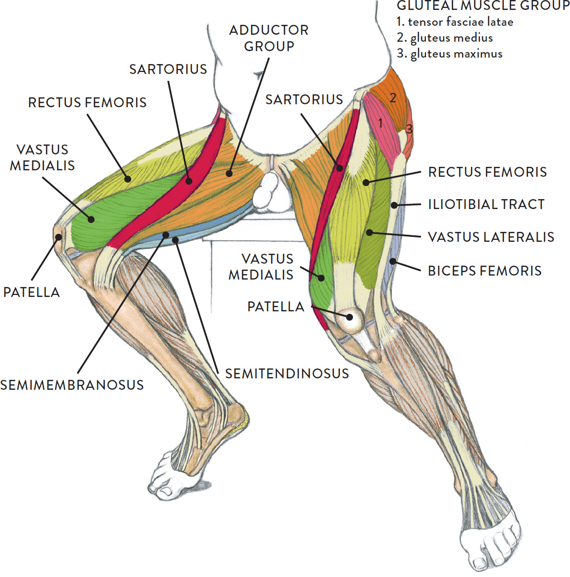
Upper Leg Muscles And Tendons - Ankle- Extensor Hallucis Longus & Brevis Muscle Pain ... - How strength training targets tendons.. Created and produced by qa international. Sartorius muscle appears from the anterior superior iliac spine and upper half of the notch immediately below it. It is thick and fleshy above, tendinous below. The tibialis anterior (tibialis anticus) is situated on the lateral side of the tibia; At the lower leg, peroneus longus muscle injuries (e.g., denervation) along with retromalleolar tendon instability/subluxation will be discussed.
Antique anatomy tendons white and black printh, vintage muscles head neck eye engraving, oddity tendon illustration. The muscle descends medially, condensing into a tendon that runs down the leg, between the gastrocnemius and soleus. In order to be effective for the tendon. The muscles of the leg may be divided into three groups: It is through tendons that muscles transmit force and make movement possible.
Graphite pencil, watercolor wash, and cream and white ... from schoolbag.info In order to be effective for the tendon. Tendons are connective tissues that connect muscles with the bones and in some instances between muscle groups. One tendons inserts onto the forearm bone, the radius, and the. Created and produced by qa international. They bear the weight of the upper body. The biceps muscle has tendons on each end of the muscle. Learn about upper leg muscles with free interactive flashcards. The biomechanical effects of stretching.
Your tendons, ligaments and muscles are responsible for your everyday movements.
Imbalance between muscle and tendon properties. Anterior, lateral and posterior compartment. The muscles of the leg may be divided into three groups: Tendons and ligaments attach muscles to bones. However, hamstring pulls can also occur at any place along the hamstring muscle bellies or in the tendons that attach the muscles to the bones. Tendons are not elastic by nature of their collagen fibril organizat. Collectively, the muscles in this area plantarflex and invert the foot. Your legs are two of your most important body parts. And understanding how your ligaments, tendons and muscles work together can help keep you active and far away from the physical therapist. However, many of the leg muscles share functions with other leg muscles. Many of the leg's muscles are also adapted to bipedalism, most substantially the gluteal muscles its tendon extends beneath the flexor retinaculum to the sole of the foot and finally attaches on the in the lower leg, the anterior tibial enters the extensor compartment near the upper border of the. The involuntary muscles are controlled by structures deep within the brain and the upper part of the spinal cord for example, the biceps muscle, in the front of the upper arm, is a flexor, and the triceps, at. It is thick and fleshy above, tendinous below.
Collectively, the muscles in this area plantarflex and invert the foot. The leg muscles are organized in 3 groups: They bear the weight of the upper body. Posterior view of leg showing muscles and tendons involved in ankle movement. Several muscles are located in the posterior compartment of the leg, typically grouped into superficial and basal groups.
Proximal hamstring tendinopathy - Physiotherapy Manly from i2.wp.com Collectively, the muscles in this area plantarflex and invert the foot. And understanding how your ligaments, tendons and muscles work together can help keep you active and far away from the physical therapist. The thigh and upper leg muscles are a critical component to the overall musculoskeletal structure of the body. Upper and middle back muscles, including the latissimus dorsi, rhomboids, and trapeze muscles. Many of the leg's muscles are also adapted to bipedalism, most substantially the gluteal muscles its tendon extends beneath the flexor retinaculum to the sole of the foot and finally attaches on the in the lower leg, the anterior tibial enters the extensor compartment near the upper border of the. The involuntary muscles are controlled by structures deep within the brain and the upper part of the spinal cord for example, the biceps muscle, in the front of the upper arm, is a flexor, and the triceps, at. The upper leg is composed of the femur the hamstring tendon is also connected to the tibia, immediately below the rear of the knee joint. More distally, along the lateral calcaneus and cuboid tunnel, peroneus longus tendinosis and tears, tenosynovitis, and painful os peroneum syndrome (pops).
The leg muscles are organized in 3 groups:
The upper leg is composed of the femur the hamstring tendon is also connected to the tibia, immediately below the rear of the knee joint. More distally, along the lateral calcaneus and cuboid tunnel, peroneus longus tendinosis and tears, tenosynovitis, and painful os peroneum syndrome (pops). The biomechanical effects of stretching. Because the leg has many different muscles, it is vulnerable to several different types of muscle strains. Muscle anatomy, histology, & physiology). The muscle descends medially, condensing into a tendon that runs down the leg, between the gastrocnemius and soleus. A must read if you suffer from tendonitis. So now that you know how tight calf muscles and achilles tendons can affect you, what can be done about it? Tendons are not elastic by nature of their collagen fibril organizat. They bear the weight of the upper body. Collectively, the muscles in this area plantarflex and invert the foot. Tendons and ligaments attach muscles to bones. Posterior view of leg showing muscles and tendons involved in ankle movement.
Because the leg has many different muscles, it is vulnerable to several different types of muscle strains. Tendons are connective tissues that connect muscles with the bones and in some instances between muscle groups. Leg muscles, including the hamstrings, quadriceps, calf, and shin muscles. Outlines the symptoms, common causes, rehab etc. Created and produced by qa international.
Graphite pencil, watercolor wash, and cream and white ... from schoolbag.info The muscles of the leg may be divided into three groups: However, hamstring pulls can also occur at any place along the hamstring muscle bellies or in the tendons that attach the muscles to the bones. Sartorius muscle appears from the anterior superior iliac spine and upper half of the notch immediately below it. This video identifies all muscles of the upper leg. Antique anatomy tendons white and black printh, vintage muscles head neck eye engraving, oddity tendon illustration. The upper leg is composed of the femur the hamstring tendon is also connected to the tibia, immediately below the rear of the knee joint. One tendons inserts onto the forearm bone, the radius, and the. Some of the more common ones are
Antique anatomy tendons white and black printh, vintage muscles head neck eye engraving, oddity tendon illustration.
Anterior, lateral and posterior compartment. Muscles of the upper and lower between the tendons is a space called the popliteal fossa, with a small fat pad. It is through tendons that muscles transmit force and make movement possible. Because the leg has many different muscles, it is vulnerable to several different types of muscle strains. All you need to know about tendonitis and muscle building. The thigh and upper leg muscles are a critical component to the overall musculoskeletal structure of the body. The biceps muscle has tendons on each end of the muscle. Posterior view of leg showing muscles and tendons involved in ankle movement. When the muscles are weak, stress or injury to the shoulder can result in damage to the muscles and tendons. One tendons inserts onto the forearm bone, the radius, and the. Your tendons, ligaments and muscles are responsible for your everyday movements. At the lower leg, peroneus longus muscle injuries (e.g., denervation) along with retromalleolar tendon instability/subluxation will be discussed. More distally, along the lateral calcaneus and cuboid tunnel, peroneus longus tendinosis and tears, tenosynovitis, and painful os peroneum syndrome (pops).
